

mri showing a slipped disc
_edited.jpg)
_edited.jpg)
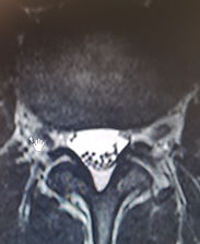
This scan shows older discs that are less well hydrated and thus appear black. The highlighted area shows that a disc has herniated or slipped into the spinal canal. This is likely to be associated with a great deal of pain in the back and leg
Before and after a discectomy


Pic1 above shows a patient with a slipped disc between L4 and L5, The obvious disc bulge is putting pressure on the L5 nerve root, Pic2 shows the appearance a year after the patient underwent a discectomy to remove the disc herniation.


A further example of a slipped disc. The picture on the left shows the disc bulging towards the spinal canal. The two smaller pics are cross sections through the disc bulge and then through a normal part of the back

In the example above, a contained disc herniation has burst causing further pain.

The disc herniation shown on the left has spontaneously resolved without surgical treatment..see also the picture below


Above, another example of a disc herniation (left pic) that has spontaneously resolved (right pic) without the need for surgical intervention

I'm a paragraph. Click here to add your own text and edit me. It's easy.
Another similar example of a disc herniation (left) that has resolved spontaneously (right)